Trauma care in emergency departments hinges upon quick and precise fracture detection to alleviate patient pain and prevent grim complications. This is where artificial intelligence (AI) has become a transformative tool that improves the diagnostic process and enables a seamless clinical workflow.
Numerous studies highlight AI's ability to detect minute fractures that may go unnoticed. For instance, a study presented at RSNA 2023 found that AI was more effective than radiologists at detecting commonly missed fractures, suggesting that a radiologist associated with AI approach could improve detection1. National standards are also beginning to acknowledge AI's role in diagnostic workflows. AI has shown utility in urgent care settings for fracture detection, as evidenced by the UK's National Institute for Health and Care Excellence (NICE). NICE endorsed Rayvolve®, which encompasses four verticals: AZtrauma, AZchest, AZmeasure, and AZboneage, for its ability to reduce diagnostic errors (nice.org.uk)2.
AI-led fracture detection is just a starting point, marking a new threshold for improving diagnostic accuracy and accelerating the patients' healthcare journey. This article examines how AI fracture detection works and its advantages for clinical settings.
Using AI for Fracture Detection in Medical Imaging
AZtrauma, fully certified MDaaS AI software for fracture detection by AZmed, uses sophisticated algorithms to analyze and detect bone fractures, dislocations, and joint effusions in medical images. Utilizing advanced deep learning and computer vision technologies, AZtrauma significantly enhances the productivity and precision of diagnosis for radiologists.
As an aid to diagnosis, AZtrauma offers several advantages over traditional diagnostics:

Advanced Machine Learning Techniques
High-quality support is provided by Convolutional Neural Networks (CNNs), visual transformers, and other advanced deep learning models, which power the software. AZmed’s Rayvolve® AI Suite is trained on one of the largest datasets of X-ray images (>15 million) with meticulous annotations by board-certified expert radiologists, enabling the AI to identify complex fracture patterns accurately. This exhaustive training transforms AZtrauma into a powerful resource. The following is an outline of how the model was developed:
- Creation of a dataset: The first step is collecting a large set of X-ray images. It includes both fractured and normal cases.
- Annotation: Expert radiologists label fractures in images, which serves as a reliable basis for training.
- Model Training: Dependent on the annotation data, the algorithms learn to distinguish fracture patterns via iterations between the two.
- Validation: Checking the accuracy and reliability of the model using new or previously untested images.
Advantages of AI Fracture Detection
AZtrauma utilizes algorithms to analyze medical images, providing improved detection of subtle fractures and lower false positives and false negatives, while also ensuring consistent readings across cases. It is this speed and accuracy that allows for the faster prioritization of critical cases, minimizing wait times and leading to on-time treatment. AZtrauma automates routine tasks, relieving radiologists of mundane workloads, allowing them to concentrate on more complex cases, reducing burnout, and improving radiology department throughput.
At RSNA 2023, Dr. Sean Raj, Chief Innovation Officer of SimonMed, presented a study validating the performance of Rayvolve®3. In this study, examination data were extracted from 2022 (without AI) and 2023 (with AI), comprising 159,601 and 170,703 examinations, respectively. The results indicated improved detection rates, with fracture prevalence of 10.4% without AI and 11.8% with AI. The mean report turnaround time (TAT) for fracture-positive cases was significantly reduced from 48 hours in the absence of AI to 8.3 hours with AI, a mean reduction of 39 hours.

Implementation in Healthcare Settings
AZmed’s approach is to provide in-depth training to healthcare professionals on basic AI principles, direct experience with AZtrauma software, and training on how to read AI-assistance diagnostic reports and how to use these insights in clinical decision-making. With different needs in infrastructure and security, AZmed offers three deployment models: On-premise & VPN, VPN, and on-premise model. Each model is designed to secure patient data and prevent data leaks, providing and managing secure network systems and controlled data storage solutions. This flexibility allows healthcare providers to implement the option that best fits their existing infrastructure and security policies.
Integration with Existing Medical Imaging Systems
AZtrauma seamlessly incorporates into hospital workflows, connecting directly into medical imaging systems for AI-powered detection:

Real-time Fracture Identification Process
When a new X-ray image is taken:
- The AI system receives the image
- The trained model analyzes the image for fracture patterns
- Results are generated within seconds, highlighting potential fractures
- Radiologists review AI-assisted findings on second capture for final interpretation
Conclusion
Using deep learning algorithms that are trained on vast datasets, AZtrauma analyzes X-rays in seconds to detect bone fractures which can sometimes be difficult to interpret. The benefits of this technology are obvious, with better diagnostic accuracy, faster results, and more potential cost-saving for health systems.
Despite the promise of AI potential in fracture detection, overcoming issues of data quality, algorithmic bias, and integration into existing healthcare systems is critical. AZmed is determined to tackle these challenges to bring AZtrauma retaining its high performance and reliability during clinical usages. With ongoing research it is expected that timely and future technology will further optimize the agent of AI for fracture diagnosis, broaden its applicability and further supplement patient care.
To learn more about the AZtrauma solution, read the clinical evidence here.
AZtrauma is now installed at SimonMed Imaging Centers. Watch the full interview conducted by JT Radiologue: RSNA 2024 Interview with SimonMed Imaging
1Erik L. Ridley. AI algorithm helps to spot overlooked fractures. RSNA. 2023
2AZmed. NHS to Use AI Technology for Faster, More Accurate Fracture Detection. AZmed website. 2024
3 Will Morton. AI cuts time for radiologists reporting fractures on x-rays. RSNA. 2023
US - Medical device Class II according to the 510K clearance. Rayvolve is a computer-assisted detection and diagnosis (CAD) software device to assist radiologists and emergency physicians in detecting fractures during the review of radiographs of the musculoskeletal system. Rayvolve is indicated for adult and pediatric population (≥ 2 years). EU - Medical Device Class IIa in Europe (CE 2797) in compliance with the Medical Device Regulation (2017/745). Rayvolve is a computer-aided diagnosis tool, intended to help radiologists and emergency physicians to detect and localize abnormalities on standard X-rays. Caution: The data mentioned are sourced from internal documents, internal studies and literature reviews. This material with associated pictures is non-contractual. It is for distribution to Health Care Professionals only and should not be relied upon by any other persons. Testimonial reflects the opinion of Health Care Professionals, not the opinion of AZmed. Carefully read the instructions for use before use. Please refer to our Privacy policy on our website. AZboneage is an uncertified feature currently under development. For more information, please contact contact@azmed.co. AZmed 10 rue d’Uzès, 75002 Paris - www.azmed.co - RCS Laval B 841 673 601© 2024 AZmed – All rights reserved. MM-25-38
Clinical Evidences
Assessing the Potential of a Deep Learning Tool to Improve Fracture Detection by Radiologists and Emergency Physicians on Extremity Radiographs
Objective
To evaluate the standalone performance of a deep learning (DL) AI based fracture detection tool on extremity radiographs and assess the performance of radiologists and emergency physicians in identifying fractures of the extremities with and without the AI aid.

Methods
The AI tool was previously developed using 132,000 appendicular skeletal radiographs divided into 87% training, 11% validation, and 2% test sets. Stand-alone performance was evaluated on 2626 de-identified radiographs from a single institution in Ohio, including at least 140 exams per body region. Consensus from three US board-certified musculoskeletal (MSK) radiologists served as ground truth. A multi-reader retrospective study was performed in which 24 readers (eight each of emergency physicians, non-MSK radiologists, and MSK radiologists) identified fractures in 186 cases during two independent sessions with and without AI aid, separated by a one-month washout period. The accuracy (area under the receiver operating curve), sensitivity, specificity, and reading time were compared with and without model aid.
Results
The model achieved a stand-alone accuracy of 0.986, sensitivity of 0.987, and specificity of 0.885, and high accuracy (> 0.95) across stratification for body part, age, gender, radiographic views, and scanner type. With AI aid, reader accuracy increased by 0.047 (95% CI: 0.034, 0.061; p = 0.004) and sensitivity significantly improved from 0.865 (95% CI: 0.848, 0.881) to 0.955 (95% CI: 0.944, 0.964). Average reading time was shortened by 7.1 s (27%) per exam. When stratified by physician type, this improvement was greater for emergency physicians and non-MSK radiologists.
Conclusion
The AI tool demonstrated high stand-alone accuracy, aided physician diagnostic accuracy, and decreased interpretation time.
Read More: Published in Academic Radiology
External validation of a commercially available deep learning algorithm for fracture detection in children
Objective
The purpose of this study was to conduct an external validation of a fracture assessment deep learning algorithm (Rayvolve®) using digital radiographs from a real-life cohort of children presenting routinely to the emergency room.

Methods
This retrospective study was conducted on 2634 radiography sets (5865 images) from 2549 children (1459 boys, 1090 girls; mean age, 8.5 ± 4.5 [SD] years; age range: 0–17 years) referred by the pediatric emergency room for trauma. For each set was recorded whether one or more fractures were found, the number of fractures, and their location found by the senior radiologists and the algorithm. Using the senior radiologist diagnosis as the standard of reference, the diagnostic performance of deep learning algorithm (Rayvolve®) was calculated via three different approaches: a detection approach (presence/absence of a fracture as a binary variable), an enumeration approach (exact number of fractures detected) and a localization approach (focusing on whether the detected fractures were correctly localized). Subgroup analyses were performed according to the presence of a cast or not, age category (0–4 vs. 5–18 years) and anatomical region.
Results
Regarding detection approach, the deep learning algorithm yielded 95.7% sensitivity (95% CI: 94.0–96.9), 91.2% specificity (95% CI: 89.8–92.5) and 92.6% accuracy (95% CI: 91.5–93.6). Regarding enumeration and localization approaches, the deep learning algorithm yielded 94.1% sensitivity (95% CI: 92.1–95.6), 88.8% specificity (95% CI: 87.3–90.2) and 90.4% accuracy (95% CI: 89.2–91.5) for both approaches. Regarding age-related subgroup analyses, the deep learning algorithm yielded greater sensitivity and negative predictive value in the 5–18-years age group than in the 0–4-years age group for the detection approach (P < 0.001 and P = 0.002) and for the enumeration and localization approaches (P = 0.012 and P = 0.028). The high negative predictive value was robust, persisting in all of the subgroup analyses, except for patients with casts (P = 0.001 for the detection approach and P < 0.001 for the enumeration and localization approaches).
Conclusion
The Rayvolve® deep learning algorithm is very reliable for detecting fractures in children, especially in those older than 4 years and without cast.
Read More: Published in Diagnostic and Interventional Imaging
Real-World Clinical Cases in X-ray Analysis with AI
Patient Case 1: Pediatric Elbow
The Rayvolve® AZtrauma module analyzed a 3-image series of pediatric elbow radiographs for AI fracture detection. The module triaged the 3 images for high suspicion and indicated the area of concern in the distal forearm, with subtle cortical buckling consistent with a torus (buckle) fracture, a pediatric-specific injury. The findings led to focused assessment from the clinician and additionally supported rapid worklist prioritization in the imaging workflow.
This case, in isolation, exemplifies how AI in fracture detection can assist in directing attention to small disruptions that may not be readily visible in complicated joints such as the elbow. AI provided pixel-level localization of the area of concern, consistently, across all 3 images, which contributed to image interpretation and would improve clinical reporting if utilized in practice.


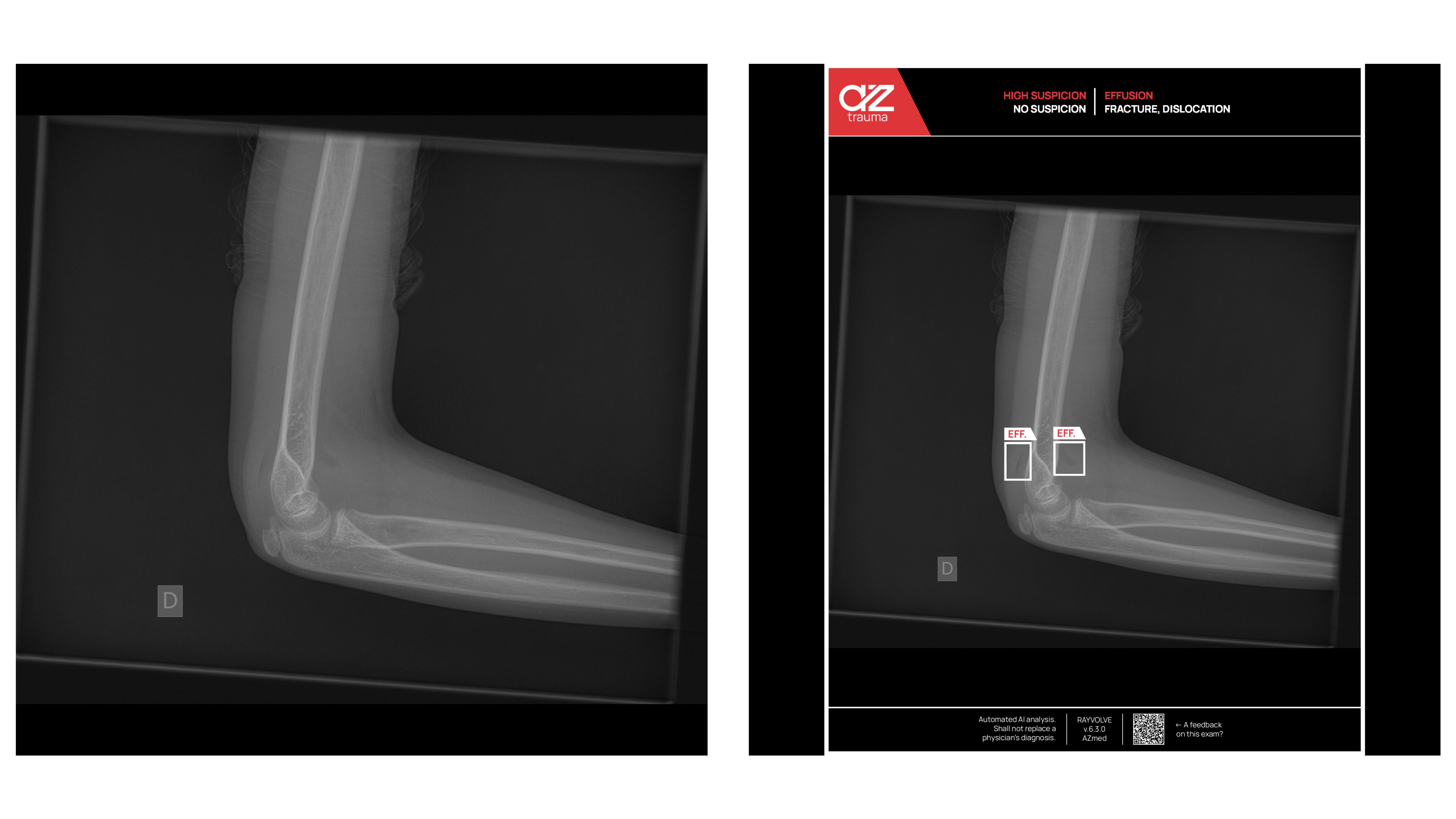
Patient Case 2: Elbow Effusion (Fat Pads)
A single-image elbow X-ray was analyzed with the use of Rayvolve® AZtrauma module, a system for fracture analysis with AI. An increased suspicion of effusion was noted and two regions of interest were highlighted, which were located in the anterior and posterior fat pads. A fat pad sign was visible which indicates the possibility of a primary intra-articular injury that may be commonly linked to occult fractures in the elbow.
The fracture analysis with AI system allowed clinicians to identify indirect signs of trauma associated with a joint effusion pattern and an abnormality that may not have shown on the plain X-ray. When indirect signs are present without a clear fracture, clinicians may complete the evaluation with CT. It is evident that AI has a role in enhancing diagnostic awareness, aiding in the interpretation of the radiograph, and supporting patient management in radiological musculoskeletal imaging.

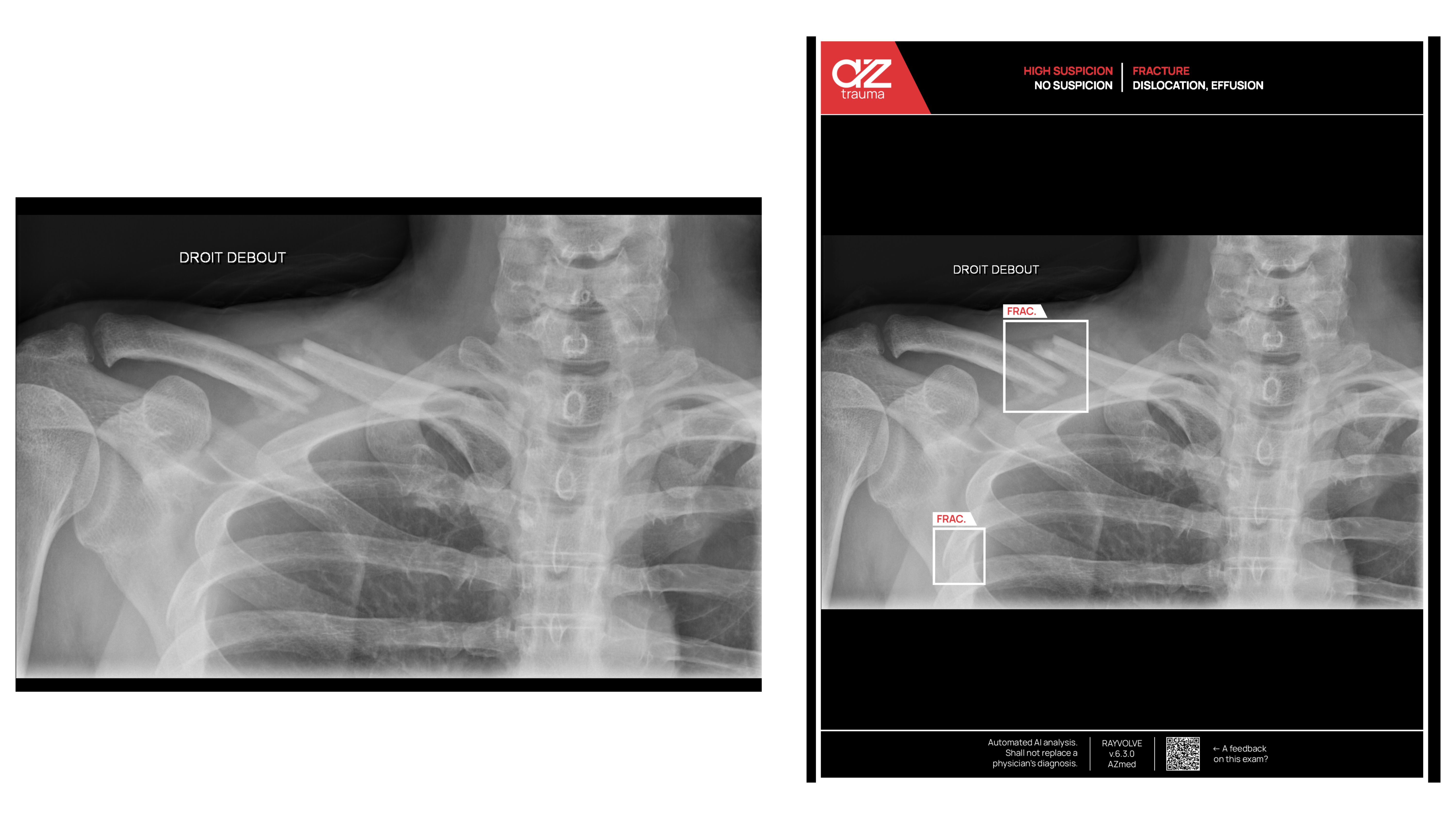
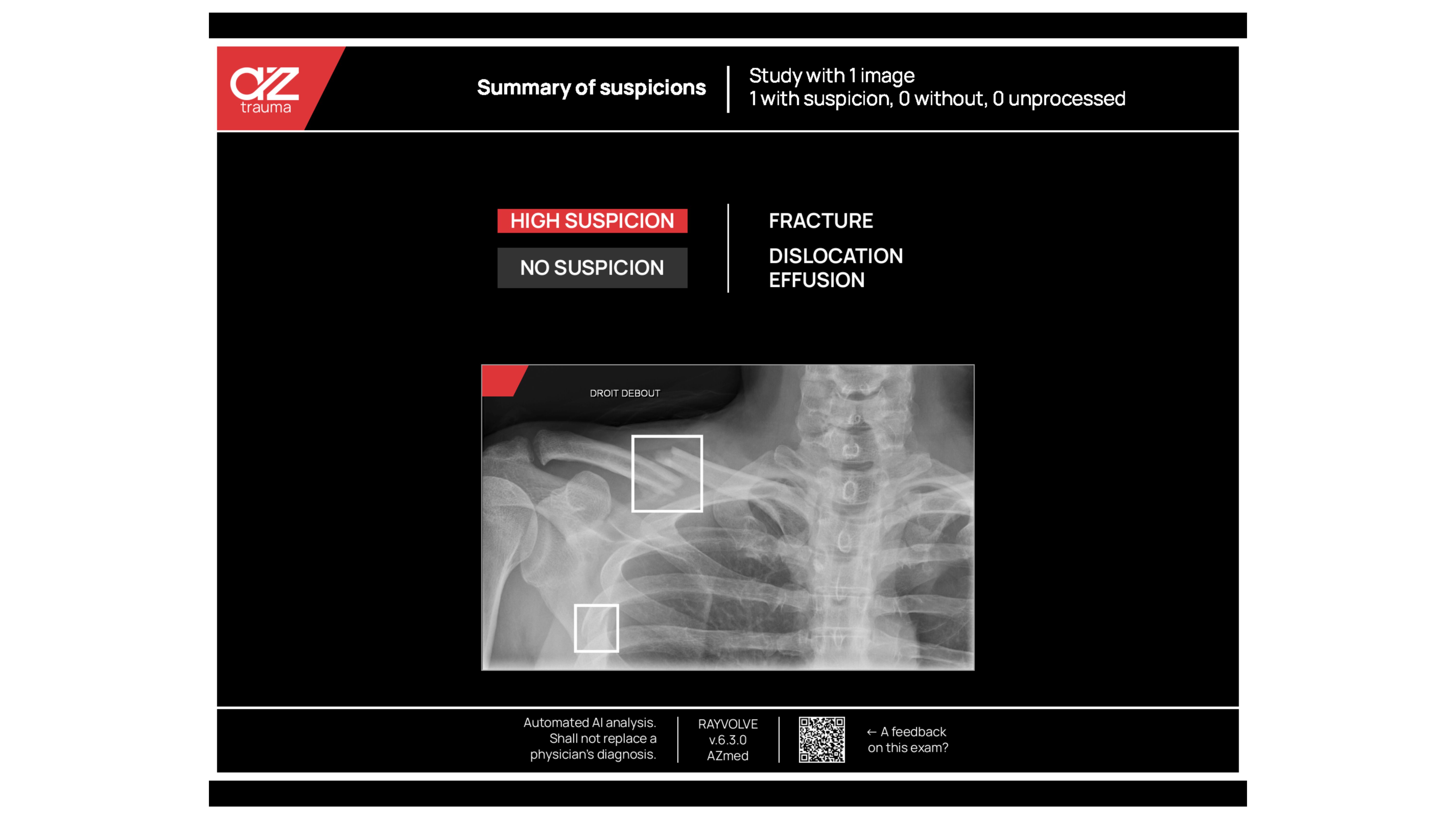
Patient Case 3: Clavicle and Rib Findings
In this instance, the Rayvolve® AZtrauma module completed an AI fracture assessment of a radiographic series, including shoulder and chest views. The system was able to identify an obvious clavicle fracture that was seen during the initial reading and also noted a rib fracture that could easily be missed due to overlapping thoracic anatomy.
The automatic localization of both lesions demonstrated that AI fracture analysis provides a “second pair of eyes” to help support diagnostic confidence, helping the radiologist be more exhaustive after detecting an obvious fracture, and mitigate the risk of missing such findings. This case illustrates the ability of AI to help radiologists to identify multiple injuries on a single study while potentially increasing accuracy and supporting reliable and consistent efficiency.
FAQs
Can AI detect fractures?
Yes, AI can detect fractures from medical images, including X-rays. Some AI-powered solutions like AZtrauma are built on top of deep learning models, which are trained on huge amounts of data to identify fractures, dislocations, and joint effusions. Studies presented at RSNA this year show that AI can detect fractures more accurately than traditional methods and can help avoid missed diagnoses.
How is AI used in radiography?
The use of AI in radiography helps radiologists by assisting in the analysis of X-ray images to detect abnormalities such as fractures. It is able to do this through the use of AI algorithms, which are trained to recognize patterns from millions of images that have been annotated, such as fractures, critical cases, and the possible diagnostic insights that can be made within seconds from Rayvolve®. This increases workflow efficiency, lowers diagnostic error rates, and aids in clinical decision-making.
Will AI replace radiologists?
No, radiologists will not be replaced by AI. Rather, it functions as an assistive tool, enhancing diagnostic certainty and easing the burden of workload, particularly in high-density clinical environments. AI is able to detect fractures and other abnormalities quickly, but the interpretations and decisions are still the domain of humans.
Is AI more accurate than radiologists?
Research has revealed that the use of AI increases the chances of detecting fractures that are likely to be missed. The studies presented at RSNA showed that AI-assisted radiologists had higher accuracy than radiologists working alone. A study from SimonMed Imaging indicated that the rate of detecting fractures increased from 10.4% (without AI) to 11.8% (with AI), and the mean report turnaround time decreased from 48 hours to 8.3 hours with the help of AI. Nevertheless, the best performance was observed when the AI was applied together with the radiologists, not in their place.
What makes AZtrauma different from other AI-powered fracture detection solutions?
AZtrauma is unique in that it is clinically validated, having received both FDA clearance and CE certification, meeting the necessary regulatory standards. It is trained on one of the biggest X-ray datasets, annotated by expert radiologists. While some AI models focus only on fractures, AZtrauma also detects dislocations and joint effusions, making it a more accurate diagnostic solution. AZtrauma is seamlessly integrated into existing medical imaging systems to ensure it works well within the radiology workflows. In real-life settings, like SimonMed Imaging Centers, it has been shown to decrease turnaround times and increase detection rates, confirming its reliability.
Which is the best AI for fracture detection on X-rays?
According to the most up-to-date head-to-head clinical evidence from Radiography Journal, AZmed’s fracture detection AI (commercially known as AZtrauma) is on par with the best tested AI tool for musculoskeletal fracture detection on X-ray, including difficult cases with metal hardware and multiple fractures.